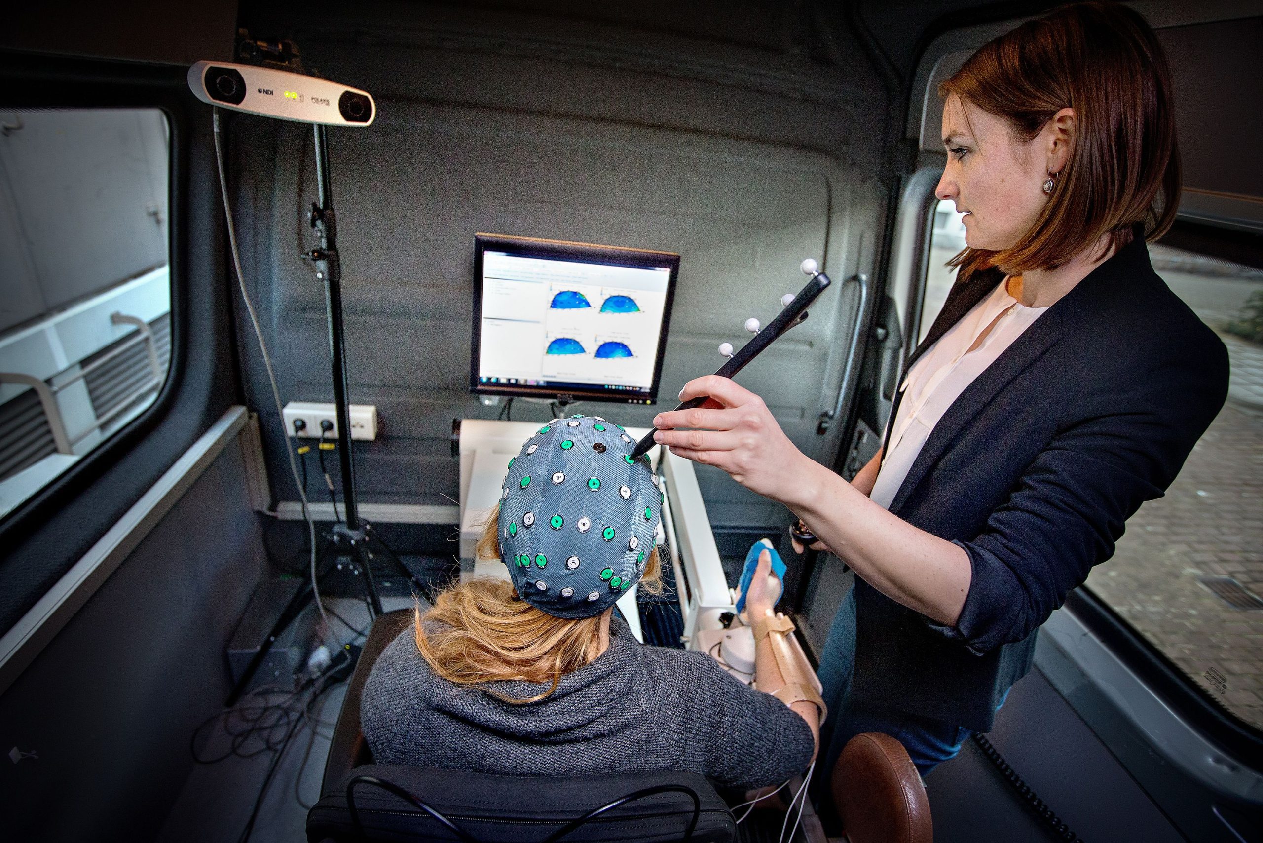
With their ‘brain neuroimaging van’ scientists from Amsterdam and Delft visit stroke patients at their home to measure brain activation patterns. By learning more about brain plasticity, they eventually hope to help improve the recovery process.
Mique Saes sits in the middle of a van parked on the campus of the VU University. The student of human movement sciences has her right arm placed in a robotic device. She feels a tingling sensation as her colleague, Lindy van Dorp, starts giving tiny electric pulses to her right index finger. Saes clenches her teeth, and hundreds of large waves appear on the electroencephalogram (EEG) on a monitor behind her, indicating muscle activity.
“During the actual measurements with patients we ask people to sit still and relax their body and not to clench their teeth in order not to disturb the EEG”, Saes explained. On her head, she wears an EEG cap with dozens of disk-shaped electrodes.
The two students demonstrated the measurements of brain activity that they normally perform on people who recently suffered a stroke. With their van, the researchers travel around the country and visit people in their houses, rehabilitation centres or nursing homes. Besides EEG, the van is equipped with a haptic robot that perturbs the arm and is used to measure the interplay between muscle and brain activity.
The project is called 4D-EEG and aims at investigating the spatial and temporal activity patterns in the brain. By performing brain measurements at intervals of several weeks for half a year, the researchers hope to gain insight into changes and adaptations of neural pathways in brains with lesions caused by stroke.
This insight could prove useful in improving the recovery process of stroke patients with motor impairment, specifically for people who have difficulties in moving their arms and hands.
TU Delft researchers developed a first prototype of the haptic robot, and later a commercial version was developed by a company called MOOG. The robot, Wristalyzer, is basically a handle that creates position and force disturbances.
“With this robot we want to better understand the relationship between motor behaviour and brain activity”, said Dr. Teodoro Solis-Escalante of the Department of Biomechanical Engineering in the Faculty of Mechanical, Maritime and Materials Engineering. “In the setup, patients have one of their arms fixed to the robot. During one of the tasks, the robot makes repetitive movements that the patients have to counteract with their wrist. On the EEG, we then see the cortical activity related to movement control.”
“The brain regions that are involved in motor control change during the recovery”, said PhD candidate Caroline Winters of the Department of Rehabilitation Medicine of the VU University Medical Center, who is also involved in the 4D-EEG project. “At first patients may use many areas of the brain, which are commonly not used. As they recover, the abnormally active brain regions diminish.”
The project is funded by the EU, by the European Research Council advanced grant worth 3.5 million euros, and runs to 2017. It is a collaboration between professor in neurorehabilitation Gert Kwakkel of VU University Medical Centre and Professor Frans van der Helm of Biomechanical Engineering, together with the Northwestern University in Chicago.
“The measurements open a new window for research that may also be applicable for neurological disorders such as Alzheimer’s disease, multiple sclerosis, and Parkinson’s Disease”, said Professor Kwakkel.


Comments are closed.